
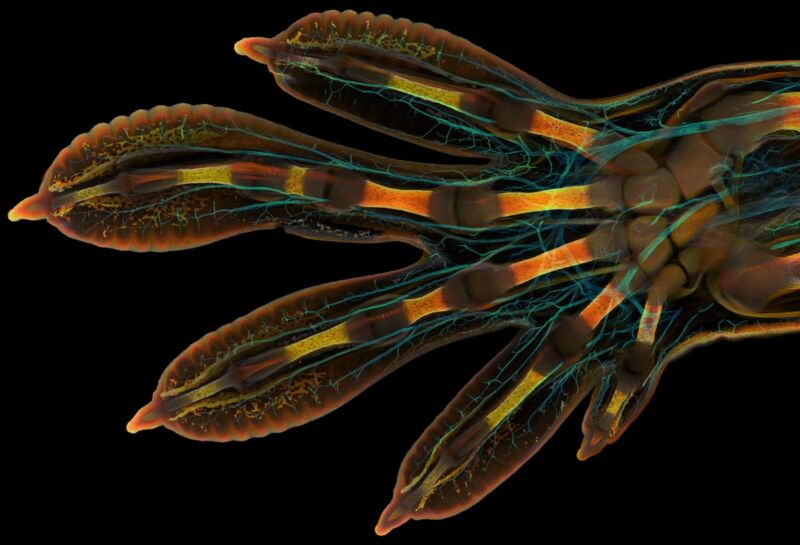
Enlarge / This arresting image of the hand of an embryonic Madagascar giant day gecko took first place in the annual competition.
The Madagascar giant day gecko (Phelsuma grandis) is a popular exotic pet, perhaps because it looks a bit like Geico's beloved animated gecko mascot. Adults measure about 10 inches in length and are known for their bright green body color, augmented by a red stripe running from the nostril to the eye. They can lick their eyeballs (a way to keep them clean since the creatures lack eyelids). And, of course, they sport those well-known adhesive pads on their feet and hands—ideal for clinging to smooth vertical surfaces—that physicists find so fascinating.
Now we have a unique perspective on the gecko's most famous appendage: a striking photomicroscopy image of an embryonic hand of Phelsuma grandis, courtesy of a Swiss graduate student, Grigorii Timin, at the University of Geneva and his advisor, Michael Milinkovitch. It's the winning image in the 2022 Nikon Small World Photomicrography Competition, designed to highlight "stunning imagery from scientists, artists, and photomicrographers of all experiences and backgrounds from across the globe," according to Nikon's communications manager Eric Flem.
The first step in creating the winning image was to prepare the sample using whole-mount fluorescent staining of the tissue. And an embryonic gecko hand is actually quite a large sample (about 3 mm or 0.12 inches long) when it comes to high-resolution microscopy. So Timin painstakingly merged hundreds of images—300 tiles, each containing some 250 optical sections—together using image-stitching to create the final result. Those cyan sections highlight the nerves in the embryonic hand, while other colors highlight bones, tendons, ligaments, skin, and blood cells.
Read 1 remaining paragraphs | Comments